

Fetal Congenital Goiter in Goat: A Case Report
Anju Kujur*, N. Srivastava, Brijesh Kumar, Nancy Jasrotia, J.Arunpandian M.
Ganeshan,
Division of Animal Reproduction, ICAR-Indian Veterinary Research Institute, Bareilly, Uttar Pradesh (243122), India.
*Email: anjikujur007@gmail.com
Abstract
A Case report of a pregnant doe aged 14 months old enabled to deliver fetus was presented in the Referral Veterinary Polyclinic, ICAR-Indian Veterinary Research Institute, Bareilly. Doe fails to deliver the foetus due to a large increament in the size of the foetus, resulting in dystocia. The doe was treated with PGF2 alpha, estrodiol and velathamate which lead to the opening of the cervix and by altering the presentation of the faulty foetus inside the uterus the dead fetus was successfully removed. The dead fetus had a large swelling in the cranio-ventral neck region. The skin was devoid of hair, pale-white and thickened with myxedema and diagnosed as congenital goitre. The most prevalent cause of congenital goitre in kids is low iodine intake or dietary iodine deficiency during the third trimester of pregnancy, which results in the birth of a dead or stillborn foetus. Iodine deficient diets, goitrogenic substances that interfere with thyroxinogenesis and excess dietary iodine are the main pathogenic pathways responsible for thyroid hyperplasia. This communication reports a case of dystocia due to congenital goiter in a goat. The defective fetus was delivered successfully by changing its presentation inside the uterus.
Keywords: Goitre, hypothyroidism, thyroid hyperplasia, Goitre Kids, dystocia
Introduction
The goitre is non-neoplastic and non–inflammatory enlargement of the thyroid. Goiter is one of the rare causes of head and neck masses in newborns, which is observed in one case per 40 000 births (Kobayashi et al., 2017). It can affect any domestic mammal, bird, or other vertebrate, although it’s most commonly seen in goat youngsters in endemic locations (Hasan et al., 2013). Congenital goitre has been reported mostly in newborn animals to dams with insufficient iodine consumption or failure to obtain dietary iodine, and is of greater economic importance than in adults due to substantial economic losses (Blood, 2000; Singh and Beigh, 2013). Iodine deficient diets-primary goitre; goitrogenic chemicals that interfere with thyroxinogenesis are the main causes of thyroid gland hypertrophy (brassica plants, soybean byproducts and water with high content of calcium and nitrates) -Secondary goitre, which can result from an excess of dietary iodine or genetically determined hereditary enzyme abnormalities in the manufacture of thyroid hormone (Radostits et al., 2007). Congenital goitre can occur due to continuous feeding of diets deficient in iodine like Subabul (Leucaenaleucocephala) (Sastry and Singh, 2008). Premature kidding also occur due to goitrogenic plant (Honparkhe et al., 2017). The main pathogenic mechanisms liable for the event of thyroid hyperplasia include iodine deficient diets, goitrogenic compounds that interfere with thyroxinogenesis, excess dietary iodine, and genetically determined defects with the enzymes liable for the biosynthesis of thyroidal hormones. These mechanisms end in inadequate thyroxine synthesis and decreased blood concentrations of thyroxine (T4) and triiodothyronine (T3) (Capen, 1995). This is often detected by the fetal hypothalamus and pituitary, stimulating a rise in thyroid stimulating hormone (TSH) production, which ends up in hypertrophy and hyperplasia of follicular cells of the thyroid. The goitre is caused by thyroid enlargement because it tries to supply the thyroid hormones needed by the animal. In utero, goitre is caused by either primary or secondary iodine deficiency (Maxi, 2007 and Kotwal et al., 2007). Most cases of congenital hypothyroidism are related to multiple late-term abortions, stillbirths, or early postnatal death. Animals born by dams being on iodine deficient diets are more likely to develop severe thyroid hyperplasia and have clinical evidence of hypothyroidism. In most cases, the sole gross lesion evident in aborted or neonatal animals may be a bilateral enlargement of the thyroid glands. Congenital goiter with alopecia and myxedema was diagnosed on the basis of gross appearance (Cheema et al., 2010; Navdeep et al., 2019). Primary goitre is caused by deficient dietary iodine intake whereas secondary goitre is caused by interference with dietary uptake of water with high content of calcium, nitrates, goitrogenic plants (Brassica sp. and a few clovers). Dystocia due to fetal goiter in doe has also been reported by (Cheema et al., 2010; Kumar et al., 2014). This is the case report of a pregnant doe giving birth to a stillborn fetus with congenital goitre which was presented in the Referral Veterinary Polyclinic, ICAR-Indian Veterinary Research Institute, Bareilly
Case history and observation
A crossbred doe aged 14 months was brought toReferral Veterinary Polyclinic, ICAR-Indian Veterinary Research Institute, Bareilly, U.P with the complaint that after completing the full term there were no signs of kidding. Per vaginal examination revealed that the cervix was not dilated; so the animal was treated for incomplete cervical dilation. The doe failed to deliver even after the treatment with PGF2 alpha and Estradiol but per vaginal examination revealed that the cervix was dilated and the fetus was without any reflex, was in anterior longitudinal presentation with severe lateral deviation of the head.
Gross examination
On gross examination, the skin of the dead kid was pale, thick with myxedema and without hair (Fig. 3). Congenital goitre with alopecia and myxedema was diagnosed on the basis of the gross picture.
Treatment
After pervaginal examination the doe was treated for incomplete cervical dilation with PGF2 alpha@250 mg, estrodiol @2-3mg,dexamethasone @ 2-5 mg and valethamate @ 10-20 mg/kg body weight intramuscularly.After 24 hours, vaginal examination revealed dilatation of the cervix withthe fetus in the anterior longitudinal presentation without any reflex. However, the doe was unable to deliver by itself. Adequate lubrication of the birth canal with liquid paraffin and gentle traction (Figure1) resulted in the successful delivery of the dead foetus (Figure2). The goat was discharged with the routine prescription of antibiotics and supportive therapy.
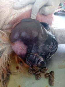
Figure1: Removal of the foetus by gentle traction
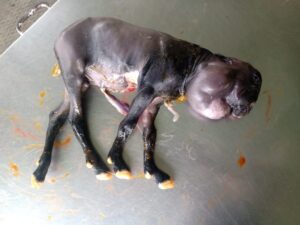
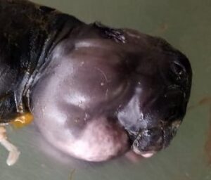
Figure 2: A dead kidFigure 3: Dead kid with alopecia and myxedema
Case discussion
Congenital goitre is a non-inflammatory and non-neoplastic enlargement of the thyroid gland in the foetus and is regarded as a common anomaly in goats (Ani et al., 1998). The condition is accompanied by an increase in the size of the foetus, myxedema, prolonged gestation and dystocia (McDonald and Pineda, 1989). Low iodine intake or failure to get dietary iodine is the common cause of congenital goiter in kids (Paulikova et al., 2002).The major pathogenic mechanisms responsible for thyroid hyperplasia include iodine deficient diets, feeding of goitrogenic compounds that interfere with thyroxinogenesis, more than required dietary iodine. These mechanisms result in inadequate thyroxine synthesis and decreased blood concentrations of (T4) and (T3). The hypothalamus and pituitary gland, stimulate an increase in (TSH) production, which results in hypertrophy and hyperplasia of follicular cells of the thyroid (Capen, 1995). Congenital hypothyroidism is associated with multiple late-term abortions, stillbirths, or early postnatal deaths (Jones et al., 1997). The kid born with Congenital goitre is generally presented with certain gross lesions like swelling on the ventral region of the neck, partial or complete alopecia of the body coat. The soft and thickened fetal skin when cut revealed myxedema. The removal of skin from the swelling within the neck revealed two massive lobes of the thyroid. These may be firm,solid and dark brown to red in colour.
Tissues from different areas of the thyroid, lungs, liver, kidneys and heart can be processed by the routine histological procedure. Tissue sections are cut at 4 µm thickness and stained by routine haematoxyline and eosin method for histopathology (Bancroft and Gamble, 2007).Thyroid tissue consists of well developed follicles of various sizes and shapes and is lined by one layer of cuboidal epithelium. Marked variation can be noted within the contents of follicles. Most of the follicles crammed with dense and dark-red colloid (thyroglobulin) and lined by flat cuboidal cells, but many follicles have a light-weight pale or dirty brownish colloid. When the follicles are depleted of thyroglobulin, the liner epithelial cells will be elongated and move towards the centre of the follicles and give a collapsed appearance. Within the lungs, the pleura and interlobular septae may also be thickened with a light-pink proteinaceous material. Similar materials are also present within the alveoli and in blood vessels. In most cases, the only gross lesion evident in aborted or neonatal animals is the bilateral enlargement of the thyroid glands. Because of the variation in the size and appearance of normal thyroid glands, histopathology must be used to confirm goitre in most cases (Capen, 1995). Ultrasonography of the thyroid gland has been frequently reported in human literature but has not been done in animals. Congenital hypothyroidism can be diagnosed during pregnancy or at birth (Mastrolia et al.,2015). Possible treatment for kids, with a deficiency of T4, is Levothyroxine sodium, which is commonly used in men, has also been studied in kids (Ommaty, 2000; Ozmen et al., 2005). Lambs with goitre have been successfully treated with 20 mg potassium iodide per os, once which is the cheapest and safest source of iodine which also give good results (Constable et al., 2017). supplementation of Lugol’s iodine in drinking water during the last month of gestation can also help the farmer to avoid iodine deficiency and such animals had normal kidding (Reddy et al., 2016). Ozmen et al., (2005) observed no congenital case of goitre when owners were advised to add potassium iodide(KI) to the feed if the dams are fed cabbage during pregnancy. Congenital goitre in a kid can successfully be treated by the use of oral administration of KI if diagnosed early.
Conclusion:
In this case enlarged thyroid glands deviated the head of the foetus, resulting in dystocia. The rotation of the foetus inside the uterus to change its presentation, however, beneficial in the delivery of the child.The congenital goitre is revealed by an enlarged thyroid with myoxedma, which is caused by dietary inadequacies or the feeding of goitrogenic chemicals in the third trimester of pregnancy. This can be avoided by putting potassium iodide in the feed and drinking lugal solution.
References
Ani, A.F.K., Khamas, W.A., Qudah, K.M.A., Rawashdah, O.A., 1998. Occurrence of congenital anomalies in Shami breed of goats; 221 cases investigated in 19 herds. Small Ruminant Research 28, 225–232.
Bancroft, J.D., Gamble, M., 2007. Theory and Practice of Histological Techniques 5th Ed, Churchill Livingstone London, UK, pp: 125–138.
Blood, D.C., Radostits, O.M., 2000. Disease caused by nutritional deficiencies. Veterinary Medicine, Bailliere- Tindall, London 1174–1177.
Capen, C.C., 1995. Endocrine system. In: Thomson’s Special Pathology. Eds: WW Carlton and MD McGavin, Missouri, Mosby. pp 250–264
Cheema, A.H., Shakoor, A., Shahzad, A.H., 2010. Congenital goitre in goats. Pakistan Vet. J. 30(1), 58–60.
Constable, P.D., Hinchcliff, K.W., Done, S.H., Grünberg, W., 2017. Veterinary Medicine.11th Edn, Elsevier Ltd, New York, USA.
Hassan, N., Randhawa, C.S., Hussian S. A., 2013. Treatment of congenital bilateral goitre in a kid-a case report. Indian Journal Science Research and Technology 1(3),19–20.
Honparkh, M., Ahuja, A.K., Dogra, P., 2017. Premature kidding due to goitrogenic plant intoxicationin beetal goat: a special case. International Journal of Science, Environment and Technology 6(4), 2303 – 2306
Jones, T.C., Hunt, R.D., King, N.W., 1997. Endocrine glands. In: Veterinary Pathology. Williams and Wilkins, Pennsylvania.pp 1236–1237
Kobayashi, M., Yagasaki, H., Saito, T., Nemoto, A., Naito, A., Sugita, K., 2017. Fetal goitrous hypothyroidism treated by intra‐amniotic levothyroxine administration: case report and review of the literature. J Pediatr Endocrinol Metab. 30(9), 1001–1005.
Kotwal, A., Priya, R., Qadeer, I., 2007. Goiter and other iodine deficiency disorders: a systematic review of epidemiological studies to deconstruct the complex web. Archives of Medical Research 38 1–14
Kumar, A., Gupta, K., Bhat, G.R., 2014. Caesarean section for treatment of fetal dystocia due to goitre in a doe. Intas Polivet 15(2), 349–350.
Mastrolia, S.A., Mandola, A., Mazor, M., 2015. Antenatal diagnosis and treatment of hypothyroid fetal goitre in an euthyroid mother: a case report and review of literature. Journal Matern Fetal Neonatal Med. 28(18), 2214–2220.
Maxi, M.G., 2007. Jubb, Kennedy and Palmer’s Pathology of Domestic Animals. 5th Edition Vol 3rd, Saunders’s Elsevier, New York, USA
McDonald, L.E., Pineda, M.H., 1989.Veterinary Endocrinology and Reproduction.4th Ed, Lea and Febiger, Philadelphia, USA.
Navdeep, S., Sethi, G.P.S., Ghuman, S.P.S., Kuldip, G., 2019 . Dystocia due to Fetal Goiter in a Goat. International.Journal.Current.Microbiology.Applied.Science. 8(01): 641–643.
Ommaty, R., 2000.Tiroidhormonpreparatlarý. In: Vademecum. Hacettepe Publishing, Istanbul. p 673.
Ozmen O., Sahinduran S., Sezer K., 2005. Clinical and pathological observations and treatment of congenital goitre in kids. Bulletin of the Veterinary Institute in Pulawy 49, 237–241.
Paulikova, I., Kovac, G., Bires, J., Paulik, S., Seidel, H., Nagy, O., 2002. Iodine toxicity in ruminants. Veterinarni Medicina, 47(12), 343–350.
Radostits, O.M., Gay, C.C., Hinchcliff, K.W., Constable, P.D., 2007. Veterinary Medicine. 10th Edition SaundersElsevier, New York, USA.
Reddy, N.V.K., Kumari, G.A., Reddy, K.R., 2016. Dystocia Due To Fetal Goiter In A Goat-A Case Report. Haryana Vet. 55 (1), 116–117.
Sastry, M.S., Singh, R., 2008. Toxic effects of subabul (Leucaena leucocephala) on the thyroid and reproduction of female goats. Indian Journal Animal Science 78, 251–253.
Singh, R., Beigh, S.A., 2013. Diseases of thyroid in animals and their management. Insights from Veterinary Medicine 9, 233–239.
https://www.pashudhanpraharee.com/causes-of-retained-placenta-in-dairy-animals/