

Successful Amputation of Forelimb in a Jersey Cross-Bred Heifer in Field Condition
A.K.Bassan1
Veterinary Assistant Surgeon ,State Veterinary Hospital, ,Department of Animal Husbandry, Nowshera, Distt. Rajouri-185151(J&K)
Abstract
The communication reports the successful surgical management of irreparable, multiple fracture of metacarpal bone by amputation, in 2.5years old, pregnant Jersey cross-bred heifer with 30 days intensive post-operative care and follow up.
Keywords: Amputation, Heifer, Fore-limb.
Introduction
Amputation is mostly indicated in cases of irreparable injuries. Comminuted open fracture with severe contamination or infection and poor blood supply is the most common reason for amputation. Non-union, ischemia, frostbite and osteomyelitis are other reasons for amputation.The cases of amputation of limb have been reported by Aheret al.,(1991) in cow; Nayak and Mohanty(1996) in buffalo; Bose et al., (2000) in leopard; Satyanarayana and Mallika (2009) in Kid.The present paper reports successful surgical management of a case of forelimb amputation in a Jersey cross-bred heiferdue to automobile accident.
Case History and Observations
2.5years old, 3 months pregnant jersey cross-bred heifer was presented to Veterinary Hospital, Nowshera, District Rajouri, J&K with the history of road traffic accident one week ago. Clinical examination revealed compound fracture of mid-shaft, right metacarpal bone, crepitation on manipulation of limb, pus discharge through the open wound, foul smell, small
fractured fragments embedded into the surrounding soft tissues (Fig 1). The edges of fractured fragments were blackish in colour and there was severe swelling around the fracture site.But, the animal was able to stand on three limbs. Further, the entire region lost its tactile and pinch reflexes. Some fracture fragments were lost due to continuous movement of limb since last one week and part below the fracture site was hanging with outer skin only. The necrosed wound emitted foul odour. After exploring the fracture site, necrosed and gangrenous muscular tissue was seen upto distal third of radius-ulna. The physical parameters of the animal were observed to be varied non-significantly. Accordingly, it was decided to perform limb amputation to save the life of the patient.
Veterinary Assistant Surgeon in Animal Husbandry Department, J&K and corresponding author
E mail : drashwanivet87@gmail.com
Surgical procedure and treatment
Sedation was achieved by administration of xylazine hydrochloride @ 0.01mg/kg body weight by i/m route. Animal was casted and restrained in left lateral recumbency with effected limb upper most. Surgical site was prepared for aseptic surgery, operative area was shaved and painted with liquid povidone-iodine. Local anaesthesia was achieved with 2% lignocaine hydrochloride by local infiltration at the site of operation. After application of tourniquet above the elbow joint to control haemorrhage, a circular skin incision was given below the elbow joint and skin flaps were reflected following blunt dissection of sub-cutaneous tissuesas described by Satyanarayana and Mallika(2009). Ligatures were applied on the muscular bundles encircling the whole circumference of limb using chromic catgut no. 2.Then the muscles were transacted below the ligatures. Bone was amputated at the level of proximal third of radius-ulna using giggly wireand articular surface was smoothen with rongeur before covering it with the flap.After completion of amputation the surgical site was lavaged using normal saline solutionwith added liquid povidone-iodine. The proximal edge of theradius enclosed within the muscular flap by applying the appositional sutures using chromic catgut no. 2. The skin defect was repaired by horizontal mattress sutures using silk thread. The suture wound was sealed with tincture benzoin seal (Fig 2).
Post-operative care and management
Post-operatively the animal was treated with Amoxicillin & Cloxacillin (inj. Intamox 3 gm, Intas Pharmaceutical, Pvt Ltd @ 15 mg/kg b.w) i/m for 5 days andInj. Meloxicam 10 ml once daily i/m route (Inj . Melonex, Intas Pharmaceutical, Pvt Ltd ) for 3 days.Regular antiseptic dressing was carried out using the liquid povidone iodine and Scavon Spray (Himalaya Pvt, Ltd) till the complete healing and sutures were removed on day12th post-operative. Proper bedding was provided to avoid risk of bedsores. The animal was kept under observation for one month and confined to stall feeding to avoid dehiscence of surgical wound and animal recovered uneventfully (Fig 3).
Conclusion
Amputation is a last option to save the life of an animal. Limb amputation is not commonly performed because of the weight of the animal and extensivepost-operativecare. There have been very few reports ofsuccessful attempt of amputation in cattle with or without prosthesis. Reports are limited mainly to heifers or light animals with the use of prosthesis. The present case reported the successful surgical management of amputation of forelimb in a Jersey CB heifer hit by automobile. Comminuted, multiple, open fracture with severe contamination, severance of blood vessels and lack of supply of blood to the dependent part might have led to necrosis of affected part which consequently demanded amputation in the present case. Young age, lower body weight of the animal and its adoption on three limbs, resulted in successful healing of the wound. Further successful amputation of limb has been reported by Suthar et al.,( 2012) as wound healing and tissue regeneration were remarkably rapid in younger animals. Animals sustaining severe internal injuries to muscles, presence of open fractures ( multiple and compound)are poor subjects for application of plaster of paris cast therefore demanded Limb amputation.
References
Aher, V.D., Sarkate, L.B., Usturge, S.M., and Bhokare, A.P., (1991). Journal of Bombay veterinary college, 3: 62.
Bose, V.S.C., Nath, I., Nayak, D.C., and Parhi, N.K., (2000). Veterinary practitioner 1:131.
Nayak, S., and Mohanty, J., (1996).Indian veterinary journal.76:42.
Satyanaryana , A., and Mallika, A., (2009).Indian veterinaryJournal .86:89.
Singh, T.B., Sharma , A.K., and Naveen Kumar.,(2003).Veterinary practitioner. 4:27.
Suthar, D.N., Patel, J.B., Modi, L.C., Patel, D.C., and Modi, F.,(2012) Indian veterinary journal.93-94.
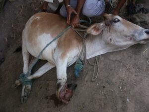
Fig 1. Compound fracture of right metacarpal bone. |
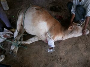
Fig 2. Immediate post operative care. |
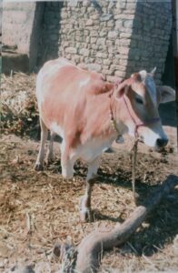
Fig 3. After one month complete healing of the wound. |
https://worldwidescience.org/topicpages/c/crossbred+holstein+friesian.html