


SURGICAL MANAGEMENT OF TEAT LACERATION IN A COW – A CASE REPORT
N.KRISHNAVENI1*
1 Assistant Professor,
Department of Veterinary Surgery and Radiology,
Veterinary College and Research Institute, Tirunelveli
Tamil Nadu Veterinary and Animal Sciences University
*corresponding author: veninarayanan110@gamil.com
Abstract
Five years old Crossbred Jersey cow was presented to the Veterinary Clinical Complex of Veterinary College and Research Institute, Tirunelveli with the history of traumatic laceration on the left fore-teat for the past 14 hours. Clinical examination revealed partial thickness deep lacerated wound on left fore-teat extending from the base to the tip with an involvement of skin and muscularis with mild swelling on the wound edges. Surgical correction of teat laceration was peformed to avoid exposing the inner structures and further infection of the teat. Surgical correction of teat laceration in cow is presented.
Key words: deep, teat, laceration, cow,
Introduction
Teat lacerations are common in cattle due to direct injuries. Teat lacerations may be superficial or deep. Superficial tat lacerations are treated with topical antiseptics and antibiotics. Deep lacerations are managed with surgical reconstruction.
History and observation
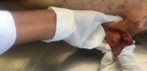
Five years old Crossbred Jersey cow was presented to the Veterinary Clinical Complex of Veterinary College and Research Institute, Tirunelveli with the history of traumatic laceration on the left fore-teat for the past 14 hours. Clinical examination revealed partial thickness deep lacerated wound on left fore-teat extending from the base to the tip of the teat with an involvement of skin and muscularis with mild swelling on the wound edges (Fig. 1). There were no leakages of milk, fistula or disruption of the mucosa. Sterile stainless steel teat siphon was inserted into the cistern of left fore-teat and milk was drained. Surgical correction of teat laceration was opted to avoid exposing the inner structures and further infection of the teat.
Treatment
Animal was restrained in a lateral recumbency and anaesthesia of teat was achieved with ring block technique using 8 ml of 2% Lignocaine HCl. Udder and teat was thoroughly cleaned with 0.1% potassium permanganate antiseptic solution and the teat was prepared aseptically for laceration repair. Surgical debridement of lacerated teat margins was performed. Partially lacerated muscularis layer was apposed using simple interrupted suture pattern with PGA 2-0. Lacerated skin edges were apposed using simple interrupted pattern with polyamaide no. 1 (Fig. 2).
Infant feeding tube no.6 was left in place for 5 days. Postoperatively Streptopenicillin 5g for 5 days and Meloxicam 150mg for 3 days was given intramuscularly. Aluspary AWD® was applied on surgical wound till suture removal. Skin sutures were removed on 10th postoperative day.
Discussion
Bovine teat is composed of five layers: Mucosa, sub mucosa, highly vascularised connective tissue muscularis and the skin (Hendrickson, 2007). Teat injuries occur due to trauma, chemical injury, insects, environmental conditions and the milking machine. The management of teat lesions will vary depending on the extent of damage to the tissue. Superficial lacerations require cleaning the wound regularly with mild antiseptic and allowing the teat to heal (Sreenu et al., 2014). Deeper lacerations with penetrating wounds require prompt response to prevent infection. Failure to repair the teat adequately leads to development of teat fistulas. Vishnugurubaran et al., 2017 reported that subcutaneous sutures with PGA and cutaneous sutures with polypropylene properly appose tissue edges and help in early healing process. Surgical intervention on the teat is best performed during first 12 hours following injury. Later swelling of the teat can be too severe to permit adequate reconstruction of the tissue (Saibaba et al., 2016). To conclude, an acute teat laceration with early surgical intervention resulted in an uneventful recovery with no complications.
Acknowledgement
The authors are thankful for the constant encouragement received from the Dean, Veterinary College and Research Institute, Tirunelveli. The authors are also thankful for the support received from Director of Clinics, TANUVAS and Associate Professor and Head, VCC, Veterinary College and Research Institute, Tirunelveli, Assistant Professor and Head, Department of Veterinary Surgery and Radiology, Veterinary College and Research Institute, Tirunelveli.
References
Hendrickson, DE. 2007. Repair of teat lacerations (Ed). In “Techniques in Large Animal Surgery” (3rd edition). Blackwell Publishing, Iowa, USA. pp 286-288.
Saibaba, M, Veena, P, Devaratnam, J, Vani, G and Suresh Kumar, R.V. 2016. Traumatic teat laceration in a Jersey cow- a case report. J Livestock sci. 7: 162-164.
Sreenu Makkena, Prakash Kumar, B, Sravanthi, P and Sudhakar Goud, K. 2014. Repair of teat laceration in a cow. Vet Clin Sci. 2 (3): pp 52-54.
Vishnugurubaran, D., Ninu, A.R. and Uma Rani, R. 2017. Surgical management of deep teat laceration in a cow. Intas Poilvet 18(2): 488-489.
 |
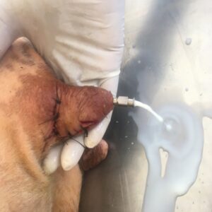 |
| Fig. 1. Left fore-teat laceration with an involvement of muscularis and skin | Fig. 2. After surgical correction of left fore-teat |