


Handling Dystocia in a Mare: A Case of Primary Uterine Inertia
Saurabh Nistane1*, Brijesh Kumar Yadav1, Pratyanshu Srivastava1, Abhishek Mishra2, Neeraj Srivastava1
1Division of Animal Reproduction, ICAR-Indian Veterinary Research Institute Izatnagar, Bareilly-243122, Uttar Pradesh, India
2Division of Veterinary Medicine, ICAR-Indian Veterinary Research Institute Izatnagar, Bareilly-243122, Uttar Pradesh, India
*Email: saurabhni99@rediffmail.com
Abstract
Dystocia in mares, though relatively uncommon, is a life-threatening condition requiring prompt intervention to ensure the survival of both the dam and foal. The present case describes the successful management of dystocia caused by primary uterine inertia in a 3-year-old primiparous mare presented with colic and restlessness. Clinical examination revealed normal fetal disposition but absence of uterine contractions, leading to the diagnosis. Supportive therapy with intravenous fluids and oxytocin infusion successfully induced uterine contractions. Subsequently, a “red bag” presentation due to premature placental separation was observed and managed immediately by rupturing the chorioallantoic membrane and applying gentle traction to deliver the foal. Post-delivery care, including antibiotics, anti-inflammatory drugs, antihistamines, and oxytocin, ensured placental expulsion and recovery of the mare. Follow-up confirmed complete recovery within 8–10 days. This case highlights that early recognition and timely intervention are critical in managing dystocia, particularly when associated with uterine inertia. Appropriate use of oxytocin, considering the mare’s sensitivity and dose-dependent response, plays a key role in successful outcomes.
Keywords– Mare, dystocia, primary uterine inertia, oxytocin.
Introduction-
Dystocia in mares refers to any interference with normal foaling caused by maternal, fetal, or placental factors. (Frazer et al., 1997; Frazer et al., 1999). A wide range of factors contribute to dystocia in mares. The most common include malposition of the head or limbs and posterior presentation of the foal. Additional causes encompass abnormal fetal posture, disproportion between foal size and the maternal birth canal, congenital malformations, failure of proper cervical dilation, pelvic fractures, and insufficient uterine contractions (Byron et al., 2003). Although rare in mares compared to other domestic species, dystocia is a critical emergency in which small mistakes can determine the survival of the mare and foal (Pynn et al., 2014; Wilkins et al., 2008).
Uterine inertia is both a potential cause and consequence of dystocia. Primary uterine inertia occurs when the uterus fails to generate sufficient contractile activity from the outset, thereby impairing fetal expulsion and delaying or preventing labor completion. Frequently cited causes of primary uterine inertia across species are uterine overextension, infection-induced degeneration of uterine muscle, hormonal irregularities during labor, calcium deficiency (Noseir et al., 2013; Threlfall et al., 2007; Roberts, 1986). It may also occur in premature parturition when hormonal interactions are not fully established. Secondary uterine inertia, also known as exhaustion inertia, characterized by the failure of the myometrium to contract effectively due to fatigue following prolonged labor ( Lu et al., 2006). Mares experiencing their first foaling are at a higher risk of dystocia than those with prior foaling history (Ball, 2005; Ginther and Williams, 1996). A prolonged first stage of labor (greater than 30 minutes) after membrane rupture indicates possible dystocia. This case documents the successful management of dystocia resulting from primary uterine inertia in a mare.
Case history and observation
A non-descript mare, approximately 3 years old and carrying her first pregnancy to term, was brought to the Referral Veterinary Polyclinic, ICAR-IVRI, Izatnagar, with complaints of colic pain and restlessness lasting 3–4 hours. During the clinical examination, the rectal temperature of the animal was recorded at 100.8˚F, poor body condition, exhibited signs of dullness and depression. Upon per- rectal examination, the foal was alive having fetal reflexes with anterior longitudinal presentation, dorso- sacral position and a normal posture. Per- vaginal examination showed cervical dilation with palpation of fetus enclosed inside fetal membranes, but no uterine contractions were detected. Consequently, dystocia due to primary uterine inertia was diagnosed.
Treatment and discussion
As the mare was dull, weak and in lateral recumbency, 1 litre of 5% dextrose via IV was administered to stabilize her condition. Then, 5 ml Tetanus toxoid administered as a deep intra-muscular injection (i/m) followed by Oxytocin (80 I.U.) as slow intravenously in 2 litres of 5% dextrose for 30 minutes. Uterine contractions began shortly afterward and half an hour later, appearance of a dark red color chorioallantoic membrane (red bag) with cervical star on its surface at vulvar opening (Fig. 1). Without much delay, soon after its appearance, a traction was applied on it with gloved lubricated hands and breaking of water bag. First, the fetal head and one forelimb were successfully brought out. The remaining forelimb was flexed and was carefully retrieved using a long hook by applying traction at the metacarpal joint (Fig. 2). Finally, the female foal was delivered with gentle manual traction.
The mare received injections as treatment: injection Melonex™ (Meloxicam) 12 ml IM for 3 days, injection Cadistin®-Vet (Chlorpheneramine Maleate) 8 ml IM for 3 days, injection Intacef® (Ceftriaxone) 3g I/V for 5 days and injection Syntocinon® (Oxytocin) 20 IU I/M resulting placenta expulsion within 30 mins. After receiving postoperative care, the animal was observed to be alert and active (Fig. 3) and was therefore discharged. Follow-up showed that the dam made a full recovery within 8–10 days.
According to the study, foals that were born alive and survived until discharge experienced a significantly shorter interval—by 13.6 minutes—between rupture of the chorioallantoic membrane and delivery, compared to foals that were either stillborn or did not survive. (Byron et al., 2003). Disturbance or anxiety during foaling can result in primary uterine inertia. This condition can be managed by minimizing stress and administering oxytocin to the dam. In normal parturition, the chorioallantois typically remains attached to the endometrium until after the foal is born. In mares, early detachment of the outer placenta can cause protrusion of the intact chorioallantois, termed “red bag” because of its dark red surface. Identification of the cervical star confirms diagnosis. Failure of membrane rupture reduces oxygen supply, leading to fetal hypoxia and possible asphyxiation if not promptly corrected.
Conclusion: Early detection of equine dystocia and quick treatment are vital for foal survival. In prolonged cases, the foal may die mainly due to placental separation and reduced oxygen supply. Different protocols for oxytocin administration include a single intramuscular dose of 40–120 IU, repeated intravenous, intramuscular, or subcutaneous doses of 5–20 IU at 15–20 minute intervals, or an intravenous infusion of 60–120 IU diluted in 1 L of saline at a rate of 1 IU per minute until the onset of second-stage labour. Since the term mare’s uterus is highly responsive to oxytocin, the effect is dose-dependent (Macpherson et al., 1997). Although uterine inertia–related dystocia is uncommon in mares, it can be effectively managed with oxytocin administration.
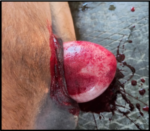
Figure 1: Red bag with cervical star
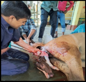
Figure 2: Traction with long hook
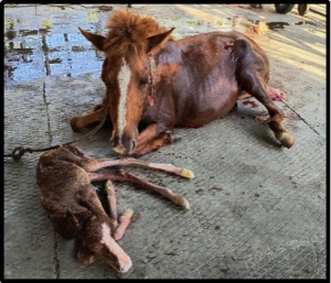
Figure 3: Mare with its live foal
References
Ball, B. A. (2005). Dystocia in the mare: Management and decision making. Proc Annu Meet Italian Assoc Equine Vet pisa p1. 4.
Byron, C. R., Embertson, R. M., Bernard, W. V., Hance, S. R., Bramlage, L. R., & Hopper, S. A. (2003). Dystocia in a referral hospital setting: approach and results. Equine Veterinary Journal. 35(1): 82-85.
Frazer, G. S., Embertson, R., & Perkins, N. R. (1997). Complications of late gestation in the mare. Equine Veterinary Education. 9(6): 306-311.
Frazer, G. S., Perkins, N. R., & Embertson, R. M. (1999). Normal parturition and evaluation of the mare in dystocia. Equine Veterinary Education. 11(1): 41-46.
Frazer, G. S., Perkins, N. R., Blanchard, T. L., ORSINl, J., & Threlfall, W. R. (1997). Prevalence of fetal maldispositions in equine referral hospital dystocias. Equine veterinary journal. 29(2): 111-116.
Ginther, O. J., & Williams, D. (1996). On-the-farm incidence and nature of equine dystocias. Journal of equine veterinary science. 16(4): 159-164.
Lu, K. G., Barr, B. S., Embertson, R., & Schaer, B. D. (2006). Dystocia—A true equine emergency. Clinical Techniques in Equine Practice. 5(2): 145-153.
Macpherson, M. L., Chaffin, M. K., Carroll, G. L., Jorgensen, J., Arrott, C., Varner, D. D., & Blanchard, T. L. (1997). Three methods of oxytocin-induced parturition and their effects on foals. Journal of the American Veterinary Medical Association. 210(6): 799-803.
Noseir, W. M. (2013). Disorders of the postpartum bovine uterus: A Literature Review. MRVSA Wael MB. Noseir. 2: 32-42.
Pynn, O. (2014). Managing mare dystocia in the field. In Practice. 36(7): 347-354.
Roberts, S. J. (1986). Dystocia. In S. J. Roberts (Ed.), Veterinary obstetrics and genital diseases (3rd ed., pp. 382–383). Woodstock, VT: Author.
THRELFALL, W. R. (2007). Retained fetal membranes. Current therapy in large animal theriogenology. 107-113.
Wilkins, P. A. (2008). Dystocia: A true emergency of the horse. In Proc Eur Equine Meeting XIV SIVE/FEEVA Congress Venice Italy p (pp. 285-289).